

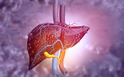
Liver cancer survival rates
Liver cancer, particularly hepatocellular carcinoma (HCC), is a highly aggressive malignancy that poses a significant global health burden. Various factors contribute to its development and progression, including chronic liver disease, viral infections, and immune dysregulation. Among the immune cells involved in liver cancer, B cells, traditionally known for their roles in antibody production and humoral immunity, have emerged as key players in tumor progression. This article explores how B cells contribute to the development and progression of liver cancer.
Liver cancer survival rates can vary depending on several factors, including the stage of cancer at diagnosis and the individual’s overall health. It is important to remember that survival rates are estimates and cannot predict individual outcomes. Here are some general statistics based on liver cancer stage:
Some stages of liver cancer survival rates–
Local stage: Cancer that is limited to the liver and has not spread to nearby lymph nodes or other organs. Localized liver cancer has a 5-year survival rate of about 31%.
Regional stage: The cancer has spread to nearby lymph nodes or tissues at this stage. Regional liver cancer has a 5-year survival rate of about 11%.
Distant stage: Distant-stage liver cancer means that cancer has spread to distant organs or tissues outside of the liver. The 5-year survival rate for distant liver cancer is approximately 3%.
It is important to note that these survival rates are averages and do not take into account individual differences or advances in treatment options. It is always advisable to consult with a healthcare professional who can provide personalized information based on your specific situation.
Inflammation and the Tumor Microenvironment
Chronic inflammation plays an important role in the development of liver cancer, creating a complex tumor microenvironment with immune cells. B cells can contribute to inflammation by releasing pro-inflammatory cytokines, such as interleukin-6 (IL-6), which promote tumor growth and angiogenesis. Additionally, B cells can interact with other immune cells, including T cells and myeloid-derived suppressor cells (MDSC), to enhance the inflammatory response and create a tumor-promoting environment.
Tumor-associated antibody response:
B cells can produce antibodies specific for tumor-associated antigens, which are proteins expressed by cancer cells. In liver cancer, B cells can produce antibodies against tumor-associated antigens, such as glypican-3 (GPC3) or alpha-fetoprotein (AFP). Although these antibodies may have potential diagnostic or therapeutic applications, they may contribute to tumor growth by promoting immune evasion and cancer cell survival.
Regulatory B cells (Bregs):
A special subset of B cells is known as regulatory B cells (Bregs). Bregs can inhibit the activity of effector T cells and natural killer (NK) cells, which weakens antitumor immunity. Studies have shown an increased presence of Bregs in liver cancer patients, suggesting their possible role in immune surveillance and tumor growth reduction.
Interactions with tumor-associated macrophages (TAMs):
B cells can interact with tumor-associated macrophages (TAMs) within the tumor microenvironment. TAMs are known to exhibit both pro-tumoral and anti-tumoral activities depending on their polarization state. B cells can influence TAM polarization toward a pro-tumoral M2-like phenotype, characterized by immunosuppressive and tumor-promoting functions. This interaction further supports the promotion of liver cancer development and progression.
The German Cancer Research Center (DKFZ) wanted to know what role gastrointestinal B cells play in the progression of nonalcoholic steatohepatitis (NASH), fibrosis, and NASH-induced HCC. They discovered that B cells promote liver cancer in two ways. Their findings have been published in the journal Hepatology.

How B Cells Boost Liver Cancer Development?
“Globally, fatty liver and NASH are assuming epidemic proportions,” says group leader Matthias Heikenwalder. B cells have two ways of producing destruction in the liver: They lead T cells to behave auto-aggressively in the small intestine via direct cell-cell communication.
Scientists replicated this in culture dishes when they paired B cells from NASH mice with CD8 T cells from a healthy animal with aggressive properties. Furthermore, IgA produced by B cells activates another type of immune cell called macrophages. These macrophages have special immunoglobulin A receptors on their surface. Activated macrophages contribute to the progression of fibrotic changes in the liver. In NASH-affected animals, blocked B cells with a specific antibody reverse both inflammation and fibrosis caused by autoreactive T cells. Heikenwalder’s team also examined tissue samples from bariatric surgery patients.
The findings are quite similar to those of mice with NASH: Tissues from NASH patients have significantly more B cells, more activated macrophages, and higher IgA levels when compared to healthy individuals. “The findings demonstrate that B cells, as well as IgA, are required to drive the pathological cascade in the development of liver cancer,” Heikenwalder concludes.
How cancer in distant organs can affect liver function?
Metastasis occurs when cancer spreads from its original site to distant organs. When cancer cells from another part of the body spread to the liver, they form new tumors. The presence of these tumors can have serious consequences for liver function. Here are some examples of how cancer in distant organs can affect liver function:
Tumor development: Tumor development in the liver can disrupt the normal structure and function of liver tissue. Tumors can compress nearby blood vessels and bile ducts as they grow in size, resulting in impaired blood and bile flow within the liver.
Liver inflammation: The presence of cancer cells in the liver can activate an immune response, resulting in inflammation. Chronic inflammation can harm liver cells and impair their normal function. This can result in hepatitis, which further impairs liver function.
Liver dysfunction: Cancer metastases can interfere with the liver’s ability to perform vital functions. The liver plays an important role in detoxification, metabolism, protein production, and nutrient storage. When cancer affects the liver, these functions can be disrupted, leading to a decline in overall liver function.
Liver enzyme abnormalities: Cancer metastases to the liver can cause abnormal levels of liver enzymes in the blood. Elevated levels of enzymes such as alanine transaminase (ALT) and aspartate transaminase (AST) are commonly observed in liver metastases, indicating liver cell damage. Impaired metabolism: The liver is responsible for metabolizing various substances including drugs, hormones,s and toxins. Liver metastases can disrupt this metabolic process, leading to changes in the clearance and processing of these substances. It can affect the effectiveness and toxicity of drugs and hormones in the body.
Jaundice: Liver metastases can block the bile ducts, causing bilirubin (a yellow pigment) to build up in the blood. This can lead to jaundice, a condition characterized by yellowing of the skin and eyes, dark urine,e, and pale stools.
Ascites: In some cases, liver metastases can cause fluid to accumulate in the abdominal cavity, a condition known as ascites. Ascites can cause abdominal swelling, discomfort, and difficulty breathing. It is important to note that specific effects on liver function may vary depending on the type of cancer and its stage. Treatment options for liver metastases may include surgery, radiation therapy, chemotherapy, targeted therapy, and immunotherapy.
Weill Cornell Medicine researchers discovered that cancer frequently releases molecules into the bloodstream that pathologically modify the liver, causing it to become inflammatory, causing fat accumulation, and disrupting its normal detoxification processes. This discovery sheds light on one of cancer’s more elusive survival strategies and opens the door to new diagnostics and therapeutics for detecting and reversing this process.
The researchers discovered that extracellular vesicles and particles (EVPs) containing fatty acids secreted by a variety of tumor types that are developing outside the liver can remotely reprogram the liver to a condition similar to fatty liver disease. According to the researchers, this mechanism was found in the livers of cancer patients and animal samples of the disease.
“Our findings show that tumors can cause significant systemic complications, including liver disease,” said study co-senior author Dr. David Lyden.
These effects are the result of specific strategies used by cancers to ensure their survival and accelerate their progression. These effects are the result of specific strategies used by cancers to ensure their survival and accelerate their progression. the researchers discovered that pancreatic cancers secrete molecules encapsulated in extracellular vesicles, which travel through the bloodstream and are taken up by the liver, preparing the organ to support the growth of new, metastatic tumors.
The new study discovered a different set of liver changes caused by distant cancer cells than they observed in animal models of bone, skin, and breast cancer that metastasize to other organs but not the liver. The study’s main finding is that these tumors accumulate fat molecules in liver cells, reprogramming the liver like obesity- and an alcohol-related condition known as fatty liver disease.
The researchers also discovered that the reprogrammed liver had higher levels of inflammation, as evidenced by higher levels of tumor necrosis factor-a (TNF-a) and lower levels of drug-metabolizing enzymes known as cytochrome P450, which break down potentially toxic molecules such as many drug molecules.
The cytochrome reduction P450 levels may explain why cancer patients become less tolerant to chemotherapy and other drugs as their disease progresses. This liver reprogramming was linked to EVPs released by distant tumors carrying fatty acids, particularly palmitic acid, according to the researchers. When fatty acid cargo is taken up by Kupffer cells, which are liver-resident immune cells, it causes the production of TNF-a, which leads to the formation of fatty liver. Despite the study’s reliance on animal cancer models, the researchers observed similar changes in the livers of newly diagnosed pancreatic cancer patients who later developed non-liver metastases.
“One of our more striking observations was that this EVP-induced fatty liver condition did not co-occur with liver metastases, suggesting that causing fatty liver and preparing the liver for metastasis are distinct strategies that cancers use to manipulate liver function,” said co-first author Dr. Gang Wang.

Hello there! Do you use Twitter? I’d like to follow you if that would be ok. I’m definitely enjoying your blog and look forward to new posts.
https://twitter.com/nusratbge. I am not active on twitter actually.
Hi my loved one I wish to say that this post is amazing nice written and include approximately all vital infos Id like to peer more posts like this
I had a great time with that, too. Despite the high quality of the visuals and the prose, you find yourself eagerly anticipating what happens next. If you decide to defend this walk, it will basically be the same every time.
Spot on with this write-up, I truly believe this site needs a lot more attention. I’ll probably be returning to read more, thanks for the information!
Hey there! I just want to give you a huge thumbs up for the great information you have got right here on this post. I am returning to your web site for more soon.
Mangaclash I just like the helpful information you provide in your articles
Jinx Manga I just like the helpful information you provide in your articles
Tech Learner I really like reading through a post that can make men and women think. Also, thank you for allowing me to comment!
FinTech ZoomUs You’re so awesome! I don’t believe I have read a single thing like that before. So great to find someone with some original thoughts on this topic. Really.. thank you for starting this up. This website is something that is needed on the internet, someone with a little originality!